To understand how back pain is produce in the body we need to mention that the spine, or vertebral column, is composed of vertebral bodies separated by intervertebral disks. There are 7 cervical vertebrae(C1-7), 12 Thoracic vertebrae(T1-12), 5 lumbar vertebrae (L1-5), 5 fused sacral vertebrae (S1-S5) and 4 fused coccygeal vertebrae. The four regions of the spine differ significantly in their flexibility.
Every pair of vertebrae of the spinal column is separated by a disc and there are more than 30 disks in the entire vertebral column. The Disk is composed of the annulus pulposus, the annulus fibrosus and the end plates. The disk is a hydraulic system that keep the vertebra apart. It acts to cushion any balance or pressure and permits the functional unit to move in flexion to the front, extension to the back, and to the side. The intervertebral discs form approximately 25 percent of the length of the vertebral column. However, this percentage varies in the different parts of the spinal column. In the cervical region the discs contribute 22 percent of the length of the column, in the thoracic region 20 percent, and in the lumbar are 33 percent.
The annulus fibrosus of the intervertebral disc is composed of 90 sheets of laminated collagen fibers that are oriented vertically in the peripheral layers and more obliquely in the central layers. The cellular elements of the disc cannot receive blood nutrients thorough the mediation of the synovial fluid but must rely on a diffusional system that provides a metabolic exchange with the vessels that lie withing the vertebral bodies. The nutrition of the intervertebral disc, at birth is performed by some blood vessels that penetrate the disc from the vertebral bone and lateral margins of the annulus, however in the adult stage the disc is totally avascular. The lumbar disc are in fact the largest avascular tissues in the body. The cells of the discs must therefore derive their nutrients and dispose of their waste metabolic products by diffusion from and to blood vessels at the disc margins.
Many of the tissued in the spine contain elastic and collagen fibers with mechanical properties. The collagen fibres in the annulus are arranged in a definitive geometric pattern, while the nucleus contains a few randomly dispersed collagen fibrils. The primary function of the intervertebral disc is to allow the spine to twist and bend to an almost continuously variable range of postures. As a resulted of their positions in the spine, the discs are subjected to compressive forces. It’s the nucleus, the most highly hydrated part of the disc, which contributes the majority of the internal pressure needed to balance the applied pressure.
The neck is the most flexible part and its discs are relatively thick, and it can be moved easily in all directions. In the chest region the discs are thinner. The lumbar region is relatively flexible because the discs in this area are thick and it has no rib cage to stiffen it. The sacral region is the least flexible part of the spine.
The intervertebral disk is a hydraulic system composed of a fibroelastic envelope containing a colloidal gel in its center. The fluid contained withing the confines of the encircling annulus is a colloidal gel and by its self-contained fluidity has all the characteristics of a hydraulic system. If increased intradiscal pressure forces fluid out of the disc, when pressure is released or decreased fluid returns into the disc by imbibition.
The disc is constructed like an automobile tire, and like a tire has a high internal pressure of its own. While a tire’s pressure comes from compressed air, the disc’s pressure is due to water. Discs are over 80 percent water. The discs’s high water content makes it highly elastic, that is, able to change its shape and then return to its original form. The disc is, in fact, one of the body’s chief shock absorbers.
Compression of the disk takes place in the annulus, if a vertebral disc unit is subjected to massive compression the fluid is rapidly squeezed out of the nucleus. The intervertebral disk has the ability to absorbe large amounts of water, and the essential compound involved in this process is a protein /Polysaccharide gel that can bind almost 90 times its own volume of water. The water is attracted to the ground substance because it contains glycosaminoglycons to high osmotic pressure to allows tissue to withstand compressive load. Osmosis leads to a high internal fluid pressure which can support a load just as the pressure of air in attire supports the weight of a car. With only a limited blood supply, especially to the inner aspects of the disc, the continual changeover of fluid in the nucleus, that it’s ability to be hydrated and dehydrated in a cyclical manner. The nucleus can balance the average compression forces when comparatively fully hydrated. When the nuclear tissue of the disc is damage this hydraulic action is lost. Normally the body weight and other compression forces are transmitted through the nucleus, the hydraulic action of which distributes pressure equally over the surface of the vertebra and the annulus fibrosus.
With nuclear destruction pressure is no longer evenly distributed and the major consequence of disc degeneration is the loss of hydrostatic properties, and the gradual decrease in the osmotic swelling pressure of the intervertebral disc. The ability of the disc to imbibe water and distributed load deteriorates with aging, largely owing to changes in the molecular proteoglycans and collagen.
When the nuclear tissue of the disc is damage this hydraulic action is lost. Normally the body weight and other compression forces are transmitted through the nucleus, the hydraulic action of which distributes pressure equally over the surface of the vertebra and the annulus fibrosus.
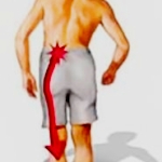
In back problems if the disc presses upon any several roots of the sciatic nerve, the person affected will feel the disctintive pain of sciatica and also may have numbness or muscular weakness in the affected leg. If the disc press instead on nerve roots in the neck, then person may feel pain shooting down an arm and have numbenesss and muscle weakness in the hand and fingers. Or if the disc presses nerves in the cauda equine, the continuation of the spinal cord supplying the bladder and bowels, the person may have trouble urinating or defecating. One common sign of back trouble is the excruciating pain called sciatica because it follows the course of the sciatic nerve, the largest nerve in the body. The sciatic nerves are formed in the hip region, one on each side, by the combination of several nerves that emerge from the lower part of the spine.
Viagra tablets uk
There are 31 pairs of spinal nerves that arise from the spinal cord at each level and exit through the intervertebral foramina. There are 8 cervical, 12 thoracic, 5 lumbar, 5 sacral, and 1 coccygeal nerve. We have 31 pairs of these nerve roots, 62 altogether, emerging from the spine. Each pair of nerves roots services a specific part of the body. The pair of nerves emerging from between the fourth and fifth neck vertebrae, for instance, activates muscles that help, over the shoulders. The nerves coming out from between the sixth and seventh neck vertebrae run to muscles serving the wrist and fingers. In the lower spine, nerves that exit from between the fourth and fifth lumbar vertebrae go to muscles moving the hips and knees, while those emerging from between the fifth lumbar vertebrae and the sacrum activate our feet.
Mechanic damage of the spine is generally believe to be a major cause of low back pain. The spine is a highly sophisticated mechanical system of which the tissues are presumably well suited to withstand the forces to which they are normally subjected. Nevertheless, if the tissues are abused, they will be damaged and act as sources of pain either directly through their own nerve supply, or indirectly, because they become distorted and compress nerve roots.
Disc lesions are probably best classified according to location, namely cervical, thoracic or lumbar, according to type, namely, bulging, protruded or herniated (ruptured); according to consistency, namely, soft or calcified, and according to position, namely midline or lateral. Bulging disks represent a very early manifestation of displacement, associated with minimal thinning of the annulus fibrosus in that particular area. They are always of soft consistency. It is at this stage that disks are probable amenable to conservative therapy. A certain degree of scarring occurs, and with bed rest, aproppiate exercises and a general improvement in overall health and posture, the weakness in the annulus fibrosus and, possibly, in the posterior longitudinal ligament often seem to disappear. In protruded disks, there is marked thinning of the annulus fibrosus, and probable few such lesions heal spontaneously, in spite of any form of conservative therapy. They maybe soft or calcified. These disks represent further progression of bulging disks and consist of lesions in which there remains merely a thin layer of annulus fibrosus overlying the protruding nucleus pulposus. Protruded disks are the most common form of disk disease.
In Intervertebral disk lesions the direction of the protrusion will determine the symptoms. The osteophytos developed and protrude in four general directions: posterior, posterolateral, lateral and anterior. Symptoms may result, depending on the anatomic structures adjacent to the vertebrae whose function is impared from compression. The ligaments that surround the disk are taut when the disk is normal, these ligaments become loose and redundant when the disk collapses. Progresive enlargement of the osteophytes may continue until they encroach on vital soft tissues adjacent to the spine, causing symptoms.
In the Cervical region the most common sites of occurrence of such lesions are between C5 and C6 and between C6 and C7, with the later being the more frequent on the two sites. In the Lumbar region the most common sites are L4 to L5 and L5 to S1 interspaces, the latter being the more frequent. Approximately 90 % of disk herniation will occur toward the bottom of the spine at L4-L5 (Lumbar segments 4 and 5) or L5-S1 (lumbar segment 5 and sacral segment 1).
Disk problems in the cervical spine will not only cause neck pain, but may experience headaches, shoulder, arm and hand pain, numbeness or weakness, chest pain, Thyroid problems, as well as ringing in the ears and blurred vision.
In the thoracic area can lead to middle back pain, pain radiating around the rib cage, chest pain, heart palpitations, difficult breathing, and headaches. And finally in the lumbar region can lead to low back pain, pain traveling down the leg, pain in the feet, bowel and bladder problems, as well as sexual organ dysfunction. In disc problems the disc don’t receive very good blood flow. Blood is responsible for carrying oxygen and nutrients to injured tissues for faster healing, and because the discs don’t receive this blood supply, then tend to be very problematic when it comes to healing.
Herniated disks, or the so-called rupture form, are seen in cases in which there is an actual tear in the annulus fibrosus and posterior longitudinal ligament, with escape of part of the nucleus pulposus through the tear. A small fragment of the nucleus pulposus may escape or there may be completed herniation, with the degenerated disk lying free.
Calcification of the end plates cartilage and vascular changes seen in older vertebrae probable impede the delivery of disc nutrients from the blood. Disc nutrition is made by diffusion, diffusion of solutes occurs through the central portion of the end plates and through the annulus. Glucose and oxygen enter via the end plates.
The nucleus pulposus may act as a chemical or a inmmunogenetic irritant to the nerve roots. The nerve roots showed different degrees of inflammatory response, which is a physical impediment to the diffusion of nutrients from the cerebrospinal fluid through the membranes of the nerve root. This produces conditions that prevent or delay the delivery of essential nutrients normally supplied by the microavasculature. Its not the mechanical pressure alone that causes the phenomena of nerve root pain but rather an abnormal chemical environment of nerve root that alters electrical activity. The nerve root can be involved in nerve root compression or nerve root irritation.
The supporting structures that give the spine stability include the anterior longitudinal ligament, the posterior longitudinal ligament, the intervertebral disks, and the musculature of the neck and trunk.


